Home » Without Label » Blood Vessels Labeled / myScience Class: The blood vessels - Elastic arteries (conducting arteries) are the largest arteries and include the aorta and other nearby branches.
Blood Vessels Labeled / myScience Class: The blood vessels - Elastic arteries (conducting arteries) are the largest arteries and include the aorta and other nearby branches.
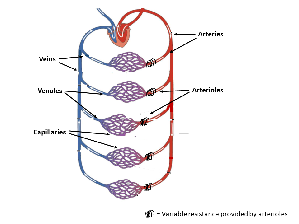
Blood Vessels Labeled / myScience Class: The blood vessels - Elastic arteries (conducting arteries) are the largest arteries and include the aorta and other nearby branches.. Veins (in blue) are the blood vessels that return blood to the heart. Capillaries come together to form venules, small blood vessels that carry blood to a vein, a larger blood vessel that returns blood to the heart. Elastic arteries (conducting arteries) are the largest arteries and include the aorta and other nearby branches. This occurs through two different circuits. The cardiovascular system consists of three kinds of blood vessels that form a closed system of passageways:
Lorenzo crumbie mbbs, bsc • reviewer: Blood travels from the heart into the subclavian artery which continues. Hma practical 3 for monday july 23 and wednesday july 25. The adventitia or outer layer which provides structural support and shape to the vessel This occurs through two different circuits.
35 Label Blood Vessels Diagram - Labels Design Ideas 2020 from upload.wikimedia.org Deep veins, located in the center of the leg near the leg bones, are enclosed by muscle. All blood vessels are basically hollow tubes with an internal space, called a lumen, through which blood flows. The three kinds of arteries are categorized by size and function: Blood vessels labeled diagram, blood vessels labeling exercises, cat blood vessels labeled, human anatomy blood vessels, human. It gives off several small branches before continuing on as the axillary artery. Arteries (in red) are the blood vessels that deliver blood to the body. The venules and veins returning blood to the heart. The common cartoid artery extends from the brachiocephalic artery.
Dr calum worsley and assoc prof craig hacking et al.
Blood vessels allow blood to circulate to all parts of the body. The basic function of these vessels is to take deoxygenated blood from different organs, supply it to the heart, and then take oxygenated blood that comes from the lungs into the heart to the rest of your body. Deoxygenated blood from the peripheral veins is transported back to the heart from capillaries, to venules, to veins, to the right side of the heart, and then. As the abdomen and pelvis contain the majority of internal organs, these regions need to be supplied by an extensive network of arteries and veins. The iliac, femoral, popliteal and tibial (calf) veins are the deep veins in the legs. Blood vessel labeling 15p image quiz. This set is often in folders with. All blood vessels are basically hollow tubes with an internal space, called a lumen, through which blood flows. Larger arteries and veins contain small blood vessels within their walls known as the vasa vasorum —literally vessels of the vessel—to provide them with this critical exchange. Blood flows throughout the body tissues in blood vessels, via bulk flow (i.e., all constituents together and in one direction). The width of blood vessels varies, but they all have a lumen. This article lists a series of labeled imaging anatomy cases by system and modality. Veins usually colored blue because oxygen poor, carry blood to the heart from the capillaries.
The function and structure of each segment of the peripheral vascular system vary depending on the organ it supplies. Blood is carried in a closed system of vessels that begins and ends at the heart the three major types of vessels are arteries, capillaries, and veins arteries carry blood away from the heart, veins carry blood toward the heart capillaries contact tissue cells and directly serve cellular needs The width of blood vessels varies, but they all have a lumen. The basic function of these vessels is to take deoxygenated blood from different organs, supply it to the heart, and then take oxygenated blood that comes from the lungs into the heart to the rest of your body. The cardiovascular system consists of three kinds of blood vessels that form a closed system of passageways:
Wire Blood Vessel Model Lower - Human Body Help from www.humanbodyhelp.com The adventitia or outer layer which provides structural support and shape to the vessel Blood vessels of the abdomen and pelvis. Elastic arteries (conducting arteries) are the largest arteries and include the aorta and other nearby branches. Veins usually colored blue because oxygen poor, carry blood to the heart from the capillaries. As the abdomen and pelvis contain the majority of internal organs, these regions need to be supplied by an extensive network of arteries and veins. The basic function of these vessels is to take deoxygenated blood from different organs, supply it to the heart, and then take oxygenated blood that comes from the lungs into the heart to the rest of your body. The systemic circuit and the pulmonary circuit (figure 20.1.1). Arteries carry oxygenated blood except in case of the pulmonary artery.
The word vascular, meaning relating to the blood vessels, is derived from the latin vas, meaning vessel.
This set is often in folders with. The iliac, femoral, popliteal and tibial (calf) veins are the deep veins in the legs. Arteries carry oxygenated blood except in case of the pulmonary artery. Name the blood vessels labeled 'e'. The function and structure of each segment of the peripheral vascular system vary depending on the organ it supplies. Aside from capillaries, blood vessels are all made of three layers: The arteries deliver freshly oxygenated blood to muscles and bone. Larger arteries and veins contain small blood vessels within their walls known as the vasa vasorum —literally vessels of the vessel—to provide them with this critical exchange. A primary purpose and significant role of the vasculature is its participation in oxygenating the body. Blood vessels allow blood to circulate to all parts of the body. Vessels transport nutrients to organs/tissues and to transport wastes away from organs/tissues in the blood. Blood vessels labeled diagram, blood vessels labeling exercises, cat blood vessels labeled, human anatomy blood vessels, human. The vessels make up two closed systems of tubes that begin and end at the heart.one system, the pulmonary vessels, transports blood from the right ventricle to the lungs and back to the left atrium.the other system, the systemic vessels, carries blood from.
The subclavian artery is the large vessel that begins the blood supply to the upper extremity. Arteries and veins transport blood in two distinct circuits: The vessels that carry blood away from the heart are called arteries, and their very small branches are arterioles. The iliac, femoral, popliteal and tibial (calf) veins are the deep veins in the legs. Human heart labeling 27p image quiz.
blood vessels - Google Search | Medical anatomy, Human ... from i.pinimg.com The three major types of blood vessels: The vessels that carry blood away from the heart are called arteries, and their very small branches are arterioles. Veins return blood back toward the heart. Larger arteries and veins contain small blood vessels within their walls known as the vasa vasorum —literally vessels of the vessel—to provide them with this critical exchange. Capillaries come together to form venules, small blood vessels that carry blood to a vein, a larger blood vessel that returns blood to the heart. The systemic circuit and the pulmonary circuit (figure 20.1.1). The three kinds of arteries are categorized by size and function: Anatomy of blood vessels review sheet 32 261 microscopic structure of the blood vessels 1.
This set is often in folders with.
Arteries (in red) are the blood vessels that deliver blood to the body. Blood vessels 11p image quiz. A primary purpose and significant role of the vasculature is its participation in oxygenating the body. The adventitia or outer layer which provides structural support and shape to the vessel The subclavian artery is the large vessel that begins the blood supply to the upper extremity. The systemic circuit and the pulmonary circuit (figure 20.1.1). Larger arteries and veins contain small blood vessels within their walls known as the vasa vasorum —literally vessels of the vessel—to provide them with this critical exchange. The three major types of blood vessels: Deoxygenated blood from the peripheral veins is transported back to the heart from capillaries, to venules, to veins, to the right side of the heart, and then. Veins return blood back toward the heart. Vessels transport nutrients to organs/tissues and to transport wastes away from organs/tissues in the blood. Arteries transport blood away from the heart. Dr calum worsley and assoc prof craig hacking et al.